

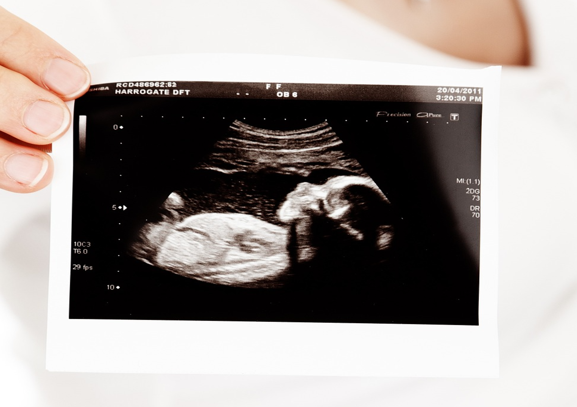
Doctors have performed one-of-a-kind brain surgery on a baby in the womb after they developed a rare malformation.
RELATED STORIES: Priyanka Chopra Reveals A Botched Nose Surgery Sent Her Into A “Deep Depression” That Nearly Derailed Her Acting Career Before It Started
The American Heart Association announced the groundbreaking procedure on Thursday, which said the surgery was done as part of a clinical trial at Boston’s Children’s Hospital and Bingham and Women’s Hospital, as reported by PEOPLE. Doctors performed the procedure to treat a vein of Galen malformation in the brain since it is a rare prenatal condition that causes the arteries to bring “high-flow, high-pressure blood to the brain from the heart.”
“Rather than to capillaries that are necessary to slow blood flow and deliver oxygen to surrounding brain tissue. Due to changes in the infant’s vascular physiology during and after the birth process, the high flow in the malformation has an even more serious effect on the heart and brain after birth, putting enormous pressure on the newborn’s heart and lungs,” the press release said.
RELATED STORIES: Toni Braxton Reveals She Underwent “Traumatic” Heart Surgery After Her Lupus Nearly Caused A Heart Attack
“This may lead to pulmonary hypertension, heart failure or other potentially life-threatening conditions. VOGM is most often first seen on a prenatal ultrasound and is definitively diagnosed by MRI during the late second or third trimester of pregnancy.”
Doctors said they discovered the malformation during a prenatal ultrasound, and an MRI diagnoses it during the later second or third trimester of pregnancy. Without treatment, the newborn can suffer from heart failure or pulmonary hypertension.
“In our ongoing clinical trial, we are using ultrasound-guided transuterine embolization to address the vein of Galen malformation before birth, and in our first treated case, we were thrilled to see that the aggressive decline usually seen after birth simply did not appear,” said lead study author Darren B. Orbach, M.D., Ph.D., co-director of the Cerebrovascular Surgery & Interventions Center at Boston Children’s Hospital.
Dr. Orbach said the infant is doing well after the surgery: “We are pleased to report that at six weeks, the infant is progressing remarkably well, on no medications, eating normally, gaining weight,”
The child’s mother gave birth two days after doctors induced her labor due to the invasive procedure.
“Due to premature rupture of membranes during the in-utero embolization, the infant was delivered by induction of vaginal birth two days later. Echocardiography after birth showed progressive normalization of cardiac output. In this case, the newborn did not require any cardiovascular support or surgery following the in-utero treatment and was watched in the NICU for several weeks after birth because of prematurity before being sent home. During that time, the newborn had a normal neurological exam and showed no strokes, fluid buildup or hemorrhage on brain MRI.”
Prenatal malformations in the brain occur during fetal development, resulting in structural or functional defects in the brain. These malformations can occur due to various factors, including genetic mutations, environmental influences, and brain development problems during pregnancy. These malformations include neural tube defects, holoprosencephaly, microcephaly, and Dandy-Walker malformation.